
- +971 (06)-882-0919
- info@tamphysioandrehab.com
- CRYSTAL PLAZA MAJAZ1 Sharjah, UAE
- +971(50)7005472

Understanding Lumbar Spinal Stenosis: Causes, Symptoms & Your Path to Relief
Explanation
Causes of Lumbar Spinal Stenosis
Spinal stenosis is primarily a degenerative condition—meaning it develops slowly over time as a result of wear and tear on the spine.
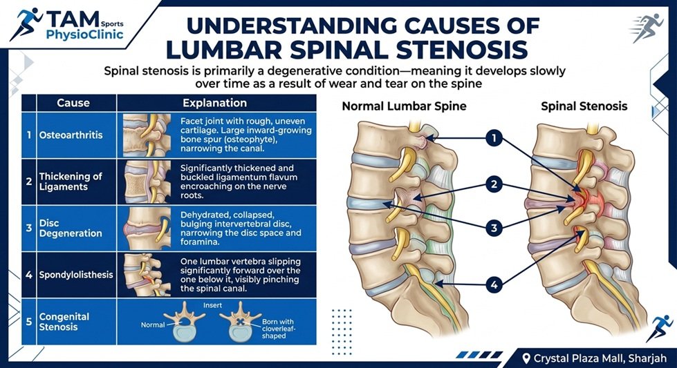
Osteoarthritis
Wear-and-tear on the facet joints (the small joints between your vertebrae) can lead to bone spurs that grow inward and narrow the spinal canal.
Thickening of Ligaments
The ligamentum flavum—a ligament that runs along the back of the spinal canal—can thicken and stiffen with age, encroaching on the nerve space.
Disc Degeneration
As discs lose height, the space between vertebrae narrows, which can reduce the openings where nerve roots exit.
Spondylolisthesis
When one vertebra slips forward over another, it can narrow the spinal canal at that level.
Congenital Stenosis
Some people are born with a naturally narrow spinal canal, making them more susceptible to symptoms as they age.
Symptoms & Clinical Picture
The hallmark symptom of lumbar spinal stenosis is
neurogenic claudication—leg pain, heaviness, or cramping that is brought on by walking or standing and relieved by sitting or leaning forward.
Common Symptoms
Pain or heaviness in the buttocks, thighs, or calves
Numbness or tingling that worsens with activity
Weakness in the legs (feeling like your legs are "giving way")
Burning sensation in the legs
Reduced walking distance before symptoms force you to stop
Relief with sitting, bending forward, or leaning on a shopping cart
The "Shopping Cart" Sign
A classic clinical picture is a patient who leans forward onto a shopping cart or a walker while walking. This forward flexion opens up the spinal canal, temporarily relieving nerve compression and allowing the patient to continue moving.
Why Does Walking Worsen Symptoms?
When you stand or walk :Gravity places a compressive load on the spine.
The spine extends (arches backward), which narrows the spinal canal.The ligamentum flavum buckles inward, further reducing available space.Increased blood flow to the nerves during activity creates a relative "demand" that cannot be met in a compressed nerve environment.Venous congestion occurs—blood pools around the compressed nerves, worsening the irritation.When you sit or lean forward, the spine flexes, which widens the spinal canal and relieves the compression.
Romberg Test
Balance with eyes closed
May be positive if proprioception is affected
Heel & Toe Walking
L5 and S1 nerve root function
May show weakness, but often less dramatic than with herniation
Flexion Test
Reproduces symptoms by arching backwar
Positive if leaning forward reduces pain
Vascular Assessment
Rules out peripheral artery disease (PAD)
Check pulses in feet to differentiate from vascular claudication
Investigations - MRI (with or without contrast)
The gold standard. Visualizes soft tissue, including ligament thickening, disc bulges, and the exact degree of canal narrowing.
CT Myelogram
An alternative if MRI is contraindicated (e.g., pacemaker). Dye is injected into the spinal fluid to show compression points.
CT Myelogram
An alternative if MRI is contraindicated (e.g., pacemaker). Dye is injected into the spinal fluid to show compression points.
X-Ray
Rules out fractures, shows alignment (spondylolisthesis), and assesses for degenerative changes.
Treatment
Conservative Management in Stages
Surgery is not the first option for most patients. Conservative management is highly effective for managing symptoms and improving function.
Surgery is not the first option for most patients. Conservative management is highly effective for managing symptoms and improving function.
Acute Symptom Management
Stage 1
- Goal: Reduce acute pain and inflammation.
- Activity Modification: Avoid prolonged standing; take frequent sitting breaks.
- Flexion Exercises: Gentle forward-bending exercises to "open up" the canal.
- Medication: NSAIDs or acetaminophen for pain.
Rehabilitation & Stabilization
Stage 2
- Goal: Improve mobility and reduce nerve irritation.
- Physical Therapy:
- Flexion-based exercises (e.g., pelvic tilts, knee-to-chest)
- Core stabilization to support the spine
- Balance and gait training
- Aquatic Therapy: Water supports body weight, allowing exercise without spinal loading.
- Assistive Devices: A walker or cane may improve walking distance.
Maintenance & Function
Stage 3
- Goal: Maintain gains and prevent progression.
- Regular Exercise Program: Continued flexion exercises, stationary cycling, swimming.
- Weight Management: Reducing body weight lessens spinal load.
- Epidural Steroid Injections: May be used to reduce inflammation around compressed nerves.
Useful Links
Subscribe Now
Don’t miss our future updates! Get Subscribed Today!