
- +971 (06)-882-0919
- info@tamphysioandrehab.com
- Crystal Plaza Mall MAJAZ 1 SHARJAH-UAE

What is a Lumbar Disc Herniation?
Understanding Lumbar Disc Herniation: Causes, Symptoms & Your Path to Recovery
To understand a herniated disc, it helps to first understand the basic anatomy of your spine. Between each of your vertebrae (the bones of your spine) sits a small, cushion-like structure called an intervertebral disc. Think of these discs as the spine's shock absorbers.
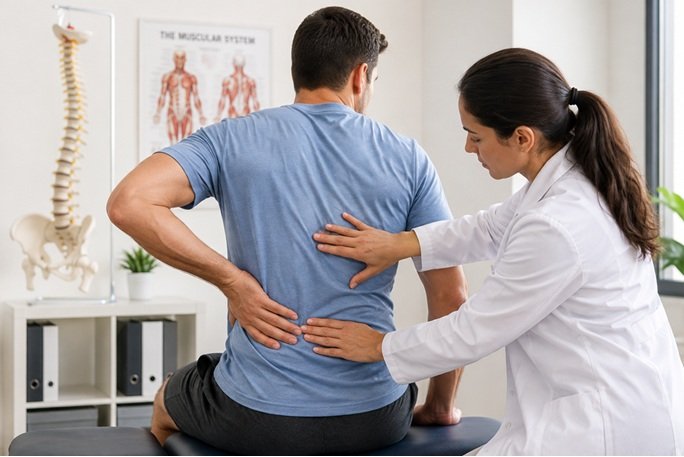
Expert guidance to help you understand your condition, identify the root cause, and find lasting relief—without unnecessary surgery.
Antomy of Intervertebral Disc
Annulus Fibrosus: A tough, tire-like outer layer. Nucleus Pulposus: A soft, jelly-like center.
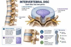
Intervertebral Disc
- Phone:+971 (50)700 5472
- Email:info@tamphysioandrehab.com
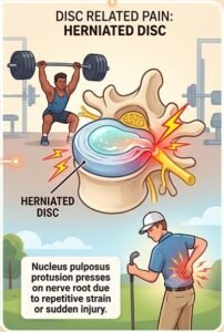
Lumbar Disc Herniation
- Phone:+971 (50) 700-5472
- Email:info@tamphysioandrehab.com
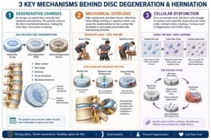
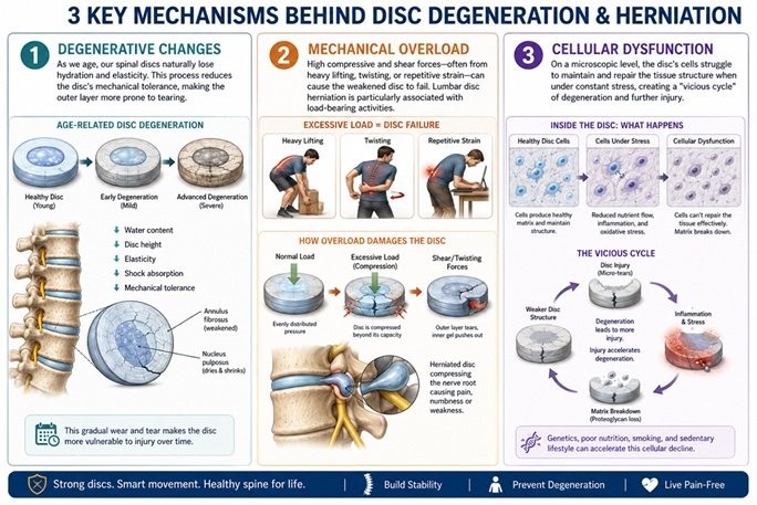
Why Does This Happen?
- Phone:+971 (50) 700-5472
- Email:info@tamphysioandrehab.com
The Clinical Picture: Symptoms and Physical Examination
Common Symptoms
The symptoms of a lumbar disc herniation depend on which nerve root is affected. The most frequently involved levels are L4-L5 and L5-S1; In a clinical study of 104 patients, 84% experienced lower back pain that radiated into the leg
Key symptoms to watch for include
Radicular Pain: A sharp, shooting, or burning pain that travels from your lower back down the buttock and into your leg. This is commonly known as sciatica.
Paresthesia: Numbness, tingling, or a "pins and needles" sensation in the leg or foot. Nearly half of patients (48%) report this
Motor Weakness: You may feel that your leg is weak, heavy, or giving way. This is due to nerve compression affecting muscle strength.
Specific Nerve Root Patterns (Disc Level: L4-L5)
The level of the herniation determines where you feel symptoms. (L4-L5)
Affected Nerve Root (L5) Pain/numbness in the lateral aspect of the thigh and leg, and the dorsum (top) of the foot. Weakness in lifting the big toe and ankle (foot drop).
Specific Nerve Root Patterns Disc Level: (L5-S1)
The level of the herniation determines where you feel symptoms. L5-S1
Affected Nerve Root :S1
Pain/numbness in the posterior (back) thigh and leg, and the sole of the foot. Weakness in pushing the foot down (plantarflexion).
Improving Lumbar Function and Stability
Massage therapy does more than mask pain—it actively improves how your spine functions. One randomized controlled study found that massage significantly improved JOA scores (a measure of lumbar function) and enhanced the function of lumbar core stabilizing muscles, including the transversus abdominis and multifidus—muscles that act as a "muscular corset" to support your spine.
Another study evaluating traditional Chinese massage (Tuina) reported that it improved posture-associated indicators, including the tone of the posterior muscle chain and lumbar posture symmetry, helping patients move and sit with better alignment
The Significance of Posture and Gait
PostureYour posture—both sitting and standing—directly affects the pressure on your discs. The L5-S1 segment, in particular, bears the brunt of this weight transfer
Sitting: The pressure on your lumbar discs is significantly higher when sitting than when standing. Slouching or "slumping" increases this pressure even further by changing the curve of your lower back.
Standing: Prolonged standing can also cause fatigue in the spinal muscles, leading to altered loading and increased stress on the lower discs -
Antalgic Posture: To reduce nerve root tension, patients often adopt a characteristic posture. In the same clinical study, 66% of patients were observed to have a scoliotic list, meaning their spine had shifted to one side in an attempt to relieve pressure on the affected nerve
Gait Analysis
How you walk provides valuable clinical clues:
Heel Walking: Difficulty walking on your heels suggests weakness in the muscles that lift your foot (ankle dorsiflexors). This is often linked to an L5 nerve root issue.
Toe Walking: Difficulty walking on your toes points to weakness in the calf muscles (plantarflexors). This is a classic sign of an S1 nerve root issue
Why Are the Muscles Failing?
Muscle weakness is not a problem with the muscle itself; it is a problem with the
Nerve signal.When a herniated disc compresses a nerve root, it blocks the electrical signals traveling from your brain and spinal cord down to your muscles. The specific muscle affected depends on the nerve root involved:L4-L5 Herniation (L5 nerve): Weakness in the Extensor Hallucis Longus (the muscle that lifts your big toe) and the Tibialis Anterior (the muscle that lifts your foot). This makes heel walking difficult
.L5-S1 Herniation (S1 nerve): Weakness in the Gastrocnemius (calf muscle). This makes toe walking difficult If the compression continues for a long time, the nerve supply is reduced, and the muscle can begin to atrophy (shrink) from disuse.
Why Does Body Weight Transfer Specifically to L5-S1?
The L5-S1 joint is the junction where the mobile spine meets the immovable pelvis. It is a region of immense physical stress and is the most common level for disc herniation.
Compressive Load: This is the area where the weight of your upper body concentrates before transferring to your legs. Studies show that the L5-S1 disc undergoes the greatest compressive deformation under sustained loading.
Reduced Shear Dissipation: The discs at this level are also less able to dissipate shear forces (forces that cause one bone to slide horizontally over another) compared to the upper lumbar segments. This reduced capacity for handling shear stress makes the L5-S1 disc more vulnerable to injury.
Sacral Base: The L5 vertebra sits on the slope of the sacrum. This angled base creates a natural shear force that the disc and joints must constantly resist, adding to the mechanical demand on this specific level.
How Does Therapeutic Massage Therapy Helps
How Massage Therapy Helps Pain ReductionMassage therapy has been shown to significantly reduce pain in patients with lumbar disc herniation. A clinical study of 100 patients found that those receiving acupoint massage achieved a 96% total effective rate in symptom improvement, compared to 82% in the control group -1. The therapy works by:Promoting local blood circulationReducing inflammation around compressed nervesReleasing endorphins (natural pain-relieving chemicals)Research on postoperative patients also demonstrates meaningful pain reduction. After just 2 weeks of treatment, patients receiving massage combined with orthopedic support saw their VAS (Visual Analogue Scale) pain scores drop from 4.34 to 2.11 for lower back pain and from 3.42 to 2.23 for leg pain
How do we assess
Special Tests for Diagnosis
Physical therapists and physicians use specific maneuvers to reproduce your symptoms and pinpoint the source of the pain.
-
Straight Leg Raise (SLR) Test / Lasègue's Test: This is the most important test for diagnosing lumbar disc herniation. You lie on your back while the clinician raises your straight leg. If this maneuver reproduces your leg pain (often at an angle between 41 and 70 degrees), it is a positive sign of nerve root irritation. In one study, 98% of patients with a confirmed disc herniation had a positive SLR test
- Crossed SLR Test (Well Test): The clinician raises the unaffected leg. If this causes pain in the affected leg, it is a strong indicator of a large, central disc herniation. This was positive in 64% of patients in the study.
- Femoral Nerve Stretch Test: This test is performed by extending the hip while the patient lies on their side. It stretches the femoral nerve (L2-L4) and is more useful for diagnosing higher-level herniations. It was positive in 40% of patients
Investigations
How Do We Confirm the Diagnosis
MRI (Magnetic Resonance Imaging): This is the gold standard for visualizing disc herniations. An MRI provides detailed images of the soft tissues, showing exactly where a disc is bulging and which nerve root it is pressing on. It is crucial for surgical planning and confirming the clinical diagnosis.
Physical Examination: While MRI is essential, it must be correlated with a thorough physical examination. In some cases, MRI findings show a disc bulge, but the patient has no symptoms. Conversely, a patient may have severe pain with minimal findings on an MRI. The combination of a careful history, physical exam, and MRI provides the most accurate diagnosis
Treatment
Conservative Management in Stages
Most patients with a lumbar disc herniation do not need surgery. Conservative (non-surgical) management is the first-line treatment and is effective in 80-90% of cases. The treatment follows a structured, stage-based approach:
Stage 1
Acute Phase (First 3–7 Days)
-
Goal: Reduce severe pain and acute inflammation.
-
Rest: Short-term bed rest (no more than 1-2 days) may be necessary for severe pain, but prolonged bed rest is discouraged as it leads to muscle wasting -8-12.
-
Medication: Non-steroidal anti-inflammatory drugs (NSAIDs) and muscle relaxants to manage pain and spasms -8.
-
Activity Modification: Avoid bending, lifting, and twisting.
Stage 2
Sub-Acute Phase (Weeks 1–6)
-
Goal: Restore pain-free range of motion and begin stabilization.
-
Physical Therapy: Begin gentle, specific exercises like the McKenzie Method or core stabilization to take pressure off the nerve.
-
Manual Therapy: Gentle mobilization or manipulation to restore joint mechanics.
-
Re-Education: Learn proper posture and body mechanics for sitting, standing, and bending.
Stage 3
Recovery & Stabilization Phase (Weeks 6–12+)
-
Goal: Strengthen supporting muscles to prevent re-injury.
-
Core Strengthening: Focus on the deep abdominal and back muscles to create a "muscular corset" that supports the spine.
-
Directional Preference Exercises: Identify and perform movements that "centralize" the pain (move the pain from your leg back into your lower back).
-
Return to Function: Gradually return to normal daily activities and work duties.
If conservative management fails after 6–12 weeks, or if there is progressive neurological weakness, treatments such as nerve blocks, steroid injections, or surgery may be considered
REACH US
Get Your Posture Assessed Today!
Buhaira Corniche Street Crystal Plaza Mall - Sharjah UAE
Our Location
Useful Links
Subscribe Now
Don’t miss our future updates! Get Subscribed Today!
©2026. TAM Physio & Rehab. All Rights Reserved.